Identification of Pythium wilt of lettuce in the field
Authors: Richard Smith, JP Dundore Arias and Steve Koike
Farm Advisor, UCCE Monterey County, Professor of Plant Pathology, California State University, Monterey Bay, and Director, TriCal Diagnostics
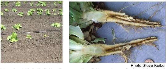
Pythium wilt, caused by Pythium uncinulatum, infects the roots of different types of lettuce plants. The pathogen survives in the soil in the form of resting spores called oospores, which are resistant to desiccation and temperature changes, and can survive in the soil for several years even in the absence of the host. Upon detecting the presence of the crop, oospores can germinate and infect plant roots directly, or form swimming zoospores, which can spread through water towards roots of the host plant. They attach to and infect the roots. Given that the pathogen needs saturated soil conditions to initiate infection, disease is often most prevalent in wetter parts of the field such as at the head or tail ends of the rows. Under severe infestations and especially with susceptible lettuce varieties, it can occur throughout the planting.
Pythium wilt has some distinctive characteristics that help in recognizing it in the field. However, there are other diseases that overlap with some of the Pythium wilt symptoms. Below is a guide for recognizing Pythium wilt in the field and to help distinguish it from other soilborne diseases. Be sure to wash the roots off to get the best view of the symptoms.
- Pythium wilt (Pythium uncinulatum):
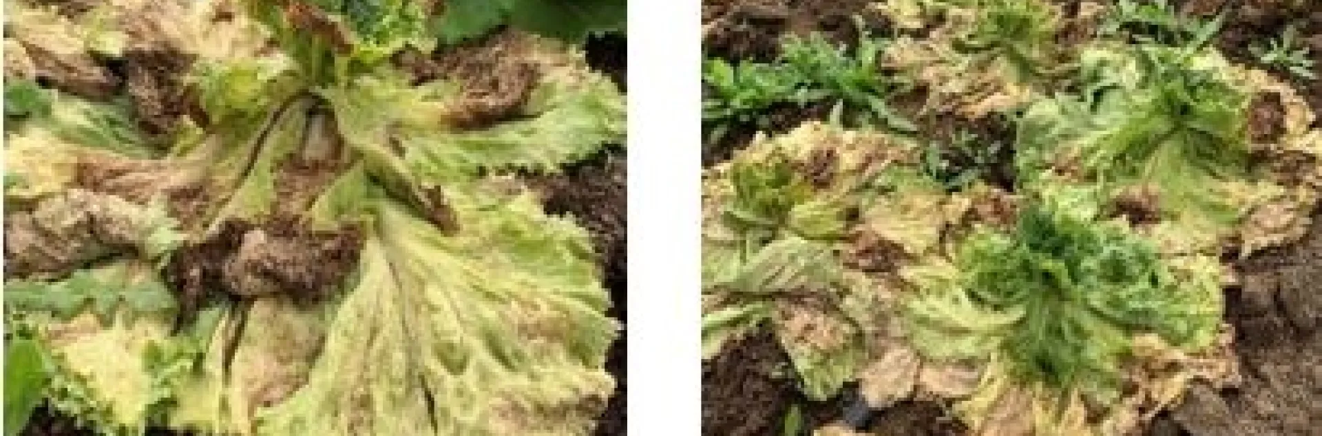
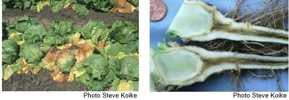
o Leaves: Older leaves wilt, turn yellow, collapse and turn brown; no distinct lesions, spots or specking (as seen with INSV); younger leaves remain upright, but in advanced stages all foliage collapses.
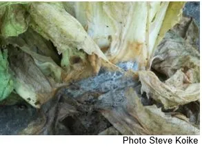
o Crowns: Do not have soft, watery decay. Mostly remain intact. Plants are not easily pulled from the ground (crowns to not readily break off).
o Roots: Taproot exterior can be distinctly dark brown to black, tissue rotted; feeder roots partially or extensively dark brown to black (discoloration does not occur in discrete bands or sections).
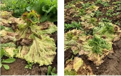
Older leaves of Pythium wilt infected plants collapse onto the ground; younger leaves often remain upright until complete death of the plant. Plants do not easily pull from ground.
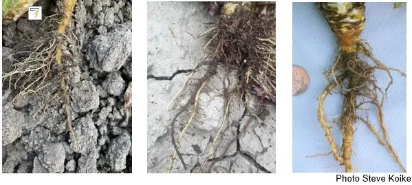
Pythium wilt: Outer tissue of the taproot (left) or feeder roots (middle and right) turn dark and are rotted.
- Lettuce Drop (Sclerotinia minor):
o Leaves: All leaves wilt, turn yellow, collapse and turn brown; no distinct lesions, spots or specking (as seen with INSV).
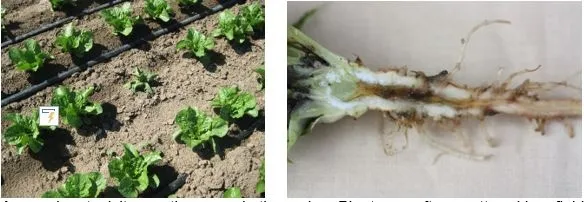
o Crowns: Have a soft, watery decay and become mushy in advanced stages; white to gray mycelium and black sclerotia (usually 1/8 inch in diameter) form on the crowns. Crowns readily break off from taproots.
o Roots: Rarely infected and therefore show no disease symptoms.
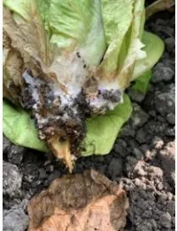
Lettuce drop infected plants have soft watery, decayed crown tissue and readily break off from the taproots when the plant is gently tugged.
- Botrytis Crown Rot (Botrytis cinerea):
o Leaves: All leaves wilt, turn yellow, collapse and turn brown; no distinct lesions, spots or specking (as seen with INSV).
o Crowns: Have a soft, watery decay and become mushy in advanced stages; crown decay is often an orange-brown color; white to gray mycelium initially forms on the crowns; later becomes gray and fuzzy. Large black sclerotia may also form on decayed crowns. Crowns readily break off from taproots.
o Roots: Are not infected and show no disease symptoms.
Botrytis crown rot attacks the crown tissue. Masses of gray mycelia are evident on infected tissue.
- Verticillium wilt (Verticillium dahliae):
o Leaves: Older leaves wilt and turn yellow. Leaf collapse occurs as plants mature and reach harvest stage.
o Crowns: Show black discoloration in the vascular tissue but not in the central core/pith.
o Roots: Taproot exterior is not discolored. Feeder roots are not discolored. Cut taproot lengthwise to show black streaking of the root vascular tissue. However, the black streaking is firm and not decayed. The vascular discoloration can be similar with symptoms caused by Fusarium wilt and ammonium toxicity.
o Other: Because plants develop symptoms near harvest, plants are rarely stunted. Head lettuce usually is the most susceptible lettuce type.
Verticillium becomes evident close to harvest. Black streaking is evident inside the tap root.
- Fusarium wilt (Fusarium oxysporum f. sp. lactucae):
o Leaves: Older leaves wilt and turn yellow; symptoms can occur on both younger and older plants.
o Crowns: Show brown to red discoloration in the vascular tissue but not in the central core/pith.
o Roots: Taproot exterior is not discolored. Feeder roots are not discolored. Cut taproot lengthwise to show brown to red streaking of the root vascular tissue. However, the brown/red streaking is firm and not decayed. The vascular discoloration can be similar with symptoms caused by Verticillium wilt and ammonium toxicity.
o Other: If infected early in development, plants may be severely stunted. If infected later in the crop cycle, plants may reach full, normal size.
Fusarium infected plants often occur in patches and are stunted with outer leaves turning yellow and necrotic. Tissue turns reddish-brown and breaks down inside the root.
- Ammonium toxicity is not a disease but is a physiological disorder. Lettuce roots are reacting to elevated levels of ammonium that build up in the soil due to cool temperatures in the spring:
o Leaves: Older leaves wilt and turn yellow; symptoms most often occur on younger plants (before and up to rosette stage).
o Crowns: Usually show no symptoms.
o Roots: Taproot exterior is not discolored. Feeder roots are not discolored. Cut taproot lengthwise to show brown to red streaking of the root vascular tissue and central root core. However, the brown/red streaking is firm and not decayed. Central pith of the root can break down and become hollow. The vascular discoloration can be similar with symptoms caused by Fusarium wilt.
o Other: Plants with ammonium toxicity damage usually are distributed randomly and singly throughout a field, in contrast with soilborne disease problems which occur in groups and patches in the field.
Ammonium toxicity mostly occurs in the spring. Plants are often scattered in a field and often, but not always, wilt in the afternoon.
These descriptions can help you to get a good idea of which disease you are dealing with. In spite of the some of the good characteristics of Pythium wilt, getting a sample to a diagnostic laboratory is the best way to be sure of which disease you are dealing with.